Mitochondrial Signaling and Metabolic Flexibility: In-Vitro Mechanisms of MOTS-c and Retatrutide
Mitochondrial dysfunction and metabolic inflexibility lie at the center of cellular aging and metabolic decline. When eukaryotic cells lose their capacity to dynamically shift between carbohydrate and lipid oxidation, metabolic efficiency drops—leading to elevated oxidative stress and impaired cellular homeostasis. In this technical deep-dive, we explore the distinct mechanisms governing mitochondrial-derived peptides like research-grade MOTS-c alongside multi-incretin GPCR agonists like HPLC-verified Retatrutide. Learn how intracellular nuclear translocation pathways differ from surface receptor signaling, review proper reconstitution mathematics, and discover co-culture paradigms for bioenergetic assays.
Mitochondrial dysfunction and metabolic inflexibility lie at the center of cellular aging, obesity, and metabolic decline. When eukaryotic cells lose their capacity to dynamically shift between carbohydrate and lipid oxidation, metabolic efficiency drops, leading to oxidative stress and impaired homeostasis.
Recent laboratory investigations have focused on mitochondrial-derived peptides and multi-receptor agonists to explore cellular bioenergetics. Among these, mitochondrial-derived peptides like research-grade MOTS-c and multi-incretin agonists such as Retatrutide serve as critical tools for understanding metabolic signaling networks.
Understanding Mitochondrial-Derived Peptides (MDPs) in Cellular Bioenergetics
Mitochondria possess their own distinct genome (mtDNA), which encodes essential components of the electron transport chain. Recent discoveries show that mitochondrial DNA also encodes short functional peptides, known as Mitochondrial-Derived Peptides (MDPs), that act as retrograde signaling molecules communicating mitochondrial status to the nucleus.
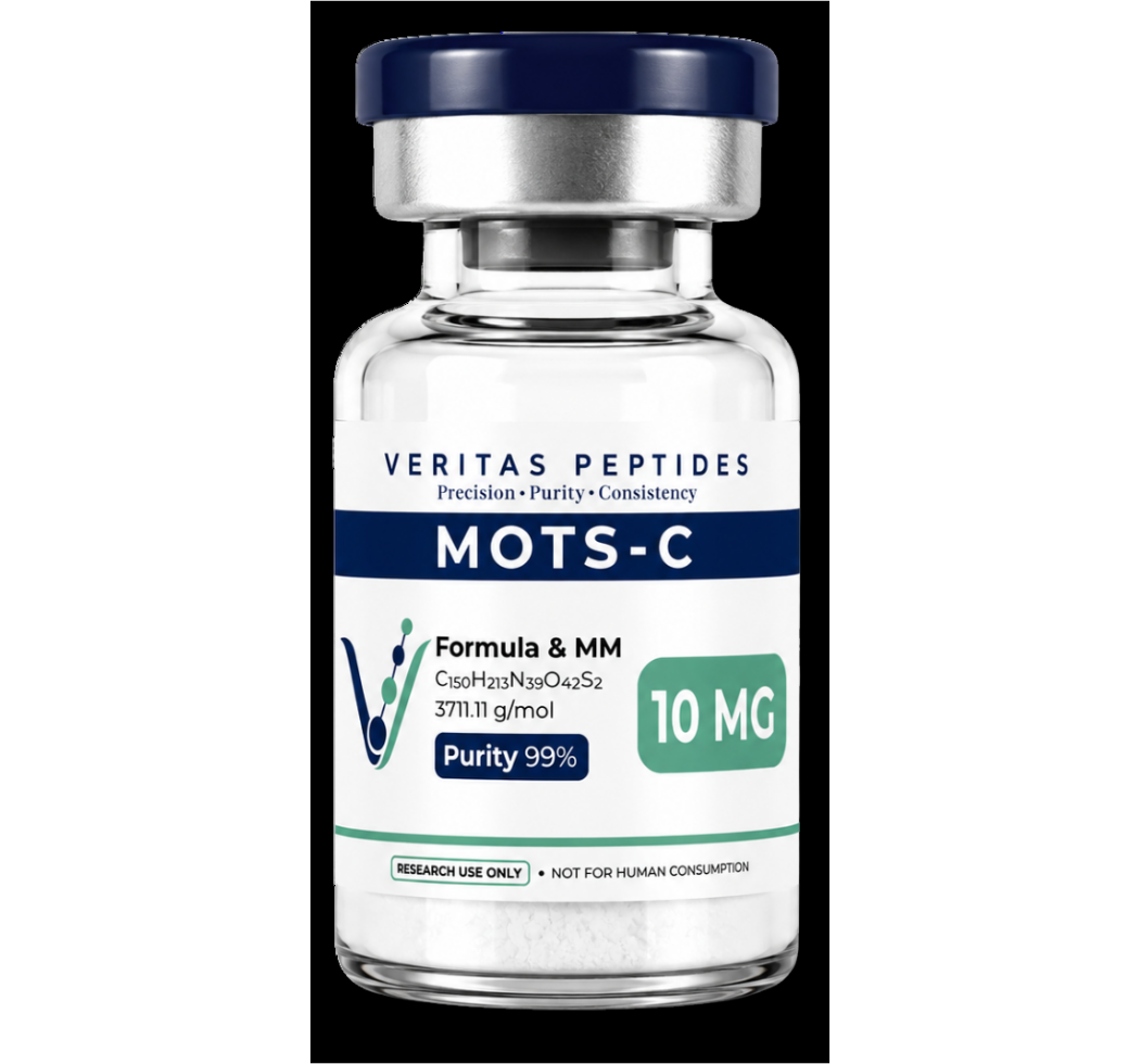
MOTS-c (Mitochondrial Open Reading Frame of the 12S rRNA Type-c) is a 16-amino-acid peptide that targets the folate cycle and purine biosynthesis pathways. Through these interactions, MOTS-c regulates intracellular energy balance via the activation of 5'-AMP-activated protein kinase (AMPK).
Primary Intracellular Cascades Triggered by MOTS-c:
- AMPK Activation: Under metabolic stress, MOTS-c translocates to the nucleus and activates AMPK, increasing the AMP-to-ATP ratio and signaling low energy status.
- Folate-Purine Interplay: By regulating the folate cycle, MOTS-c reduces cellular accumulation of 5-aminoimidazole-4-carboxamide ribonucleotide (AICAR), indirectly boosting native AMPK phosphorylation.
- Enhancement of Insulin Sensitivity: In-vitro models demonstrate that cell lines cultured with MOTS-c exhibit upregulated GLUT4 transporter translocation, enhancing glucose clearance independent of classical insulin pathways.
- Fatty Acid Oxidation: Activation of the AMPK-ACC pathway leads to down-regulation of acetyl-CoA carboxylase (ACC), lifting the inhibition on carnitine palmitoyltransferase-1 (CPT-1) and driving mitochondrial beta-oxidation.
Scientists examining cellular respiration parameters can cross-reference kinetic data in our open-access Citation Library for documented concentration-response curves.
Triple-Agonism and Metabolic Receptors: The Mechanics of Retatrutide
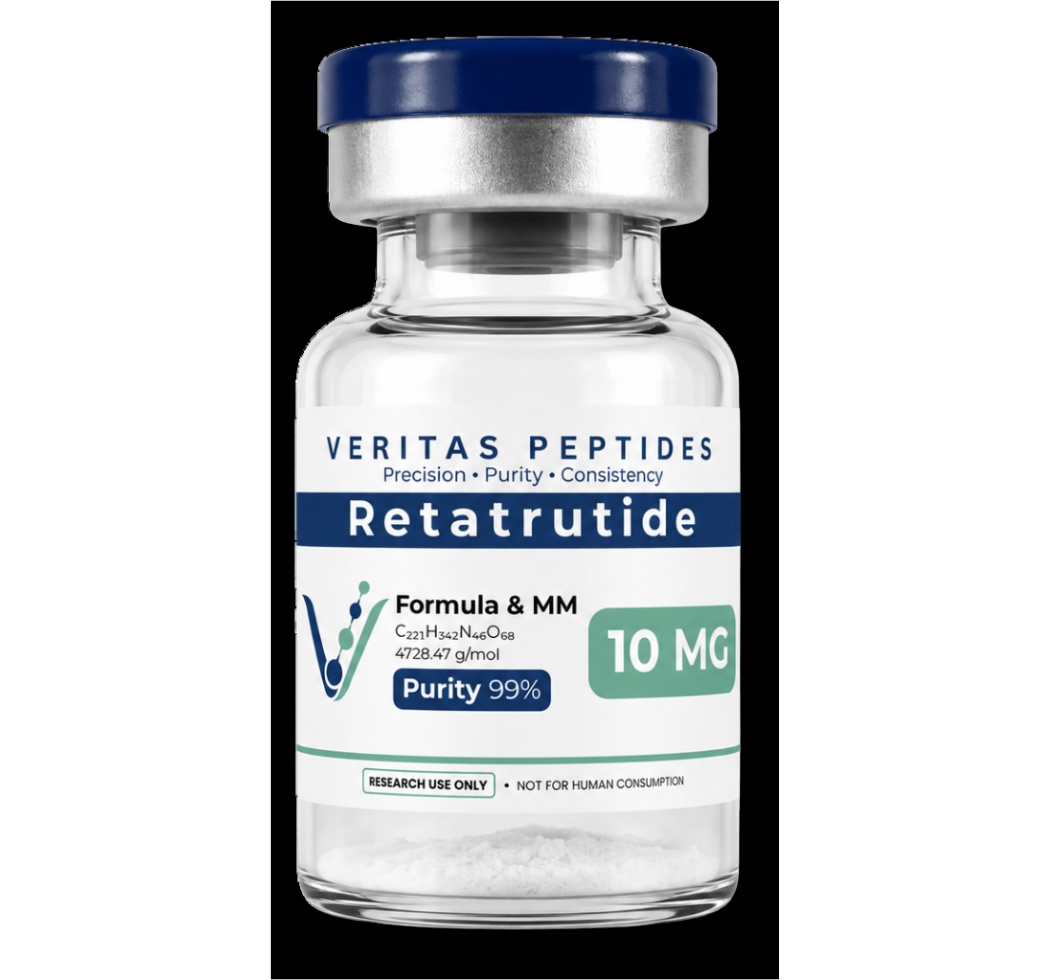
While MDPs modulate intracellular signaling directly, peptide-based multi-receptor agonists target membrane-bound G-protein coupled receptors (GPCRs) to alter downstream cellular behavior. Retatrutide represents a modern triple agonist, engineered to engage three key metabolic receptors:
- GLP-1 (Glucagon-Like Peptide-1 Receptor): Stimulates glucose-dependent insulin secretion and modulates satiety cascades.
- GIP (Glucose-Dependent Insulinotropic Polypeptide Receptor): Enhances beta-cell function and regulates lipid deposition pathways.
- GCGR (Glucagon Receptor): Increases energy expenditure and accelerates hepatic lipid catabolism.
Synergistic Signaling Dynamics
The simultaneous activation of GLP-1, GIP, and Glucagon receptors creates a distinct metabolic phenotype in cell models. While GLP-1 and GIP activation promotes insulin secretion and glucose uptake, GCGR activation elevates cyclic AMP (cAMP) levels in hepatocytes, stimulating glycogenolysis and mitochondrial fatty acid oxidation.
When evaluating multi-receptor agonists, researchers must maintain consistent peptide integrity. To ensure compound purity, review our lot-specific COA data prior to culture administration.
Comparing MOTS-c and Retatrutide: Distinct Molecular Targets
Understanding the distinction between intracellular mitochondrial signaling and extracellular GPCR activation is critical for designing targeted in-vitro models.
- Primary Site of Action: MOTS-c acts internally via nuclear translocation and AMPK activation, whereas Retatrutide binds surface GPCRs (GLP-1R, GIPR, GCGR).
- Primary Metabolic Driver: MOTS-c drives folate cycle modulation and direct mitochondrial gene transcription, while Retatrutide drives cAMP-mediated intracellular signaling cascades.
- Subcellular Effect: MOTS-c enhances autonomous mitochondrial biogenesis and insulin-independent glucose transport, whereas Retatrutide coordinates systemic hormonal signaling and substrate utilization.
- In-Vitro Application Focus: MOTS-c is ideally suited for cellular aging, metabolic stress, and mitochondrial dysfunction assays. Retatrutide provides a robust model for multi-receptor cross-talk, incretin dynamics, and hepatic lipid clearance studies.
Reconstitution Protocols and Concentration Calculations for In-Vitro Assays
Accurate reconstitution is essential for reproducible cellular assays. Synthetic peptides must be dissolved in sterile, pH-stable media or water to maintain structural stability.
Laboratory Reconstitution Workflow:
- Equilibrate lyophilized peptide vials to room temperature before reconstitution to prevent moisture condensation.
- Clean the rubber stopper using a sterile alcohol pad.
- Draw the targeted volume of sterile diluent, such as high-purity Bacteriostatic Water.
- Direct the diluent stream against the glass wall of the vial, allowing the solvent to submerge the lyophilized cake.
- Swirl gently until fully dissolved. Avoid vigorous shaking, which can cause mechanical shear stress and peptide denaturation.
Calculating Reconstitution Concentrations
Mathematical calculations for lab dilutions rely on straightforward linear equations:
- Final Concentration (mg/mL) = Mass of Peptide (mg) / Volume of Diluent (mL)
- Target Dose Volume (mL) = Target Mass (mg) / Final Concentration (mg/mL)
For rapid laboratory verification, researchers can utilize our interactive peptide reconstitution calculator to eliminate concentration errors.
Advanced Co-Culture Paradigms: Dual-Targeting Bioenergetics
In contemporary bioenergetic studies, researchers frequently explore dual-pathway dynamics. By pairing an intracellular mitochondrial activator like MOTS-c with membrane-bound signaling peptides, such as high-purity BPC-157 for tissue repair models or growth factor secretagogues like research-grade Sermorelin and HPLC-tested Tesamorelin, laboratories can map complex metabolic networks across diverse cellular environments.
Frequently Asked Questions (FAQ)
What is the primary mechanism by which MOTS-c alters cellular metabolism?
MOTS-c translocates to the cell nucleus during metabolic stress and regulates the folate-purine synthesis pathway. This triggers downstream activation of AMPK, promoting glucose uptake, enhancing fatty acid oxidation, and maintaining cellular homeostasis.
How does Retatrutide differ from traditional single or dual incretin agonists?
Retatrutide incorporates glucagon receptor (GCGR) agonism alongside GLP-1 and GIP receptor activity. The addition of GCGR signaling directly increases cellular energy expenditure and lipid metabolism beyond the scope of mono- or dual-agonist peptides.
Why is Bacteriostatic Water preferred over Sterile Water for multi-dose peptide vials?
Bacteriostatic Water contains 0.9% benzyl alcohol, which inhibits microbial growth and preserves solution sterility across multiple sampling withdrawals during extended laboratory protocols.
For laboratory and research use only. Not for human or veterinary use.